Reproductive Microsurgery
Sometimes Less is More
Microsurgery is surgery is performed with an underlying philosophy of delicate handling of the tissue, a commitment to maintaining normal architecture, minimum to no tissue trauma, and most importantly, preserving the woman's childbearing potential. Microsurgical techniques involve gentle handling of the tissue, keeping the surrounding tissues moist (dry tissue becomes damaged tissue and damaged tissue heals with scarring), use of smaller and less-reactive sutures, minimizing blood loss, and avoidance of tissue abrasion/irritation from surgical sponges, powdered gloves, and lint. A microsurgeon upholds the philosophy that maintaining and/or restoring normal architecture and childbearing potential is the primary goal and that removing the pathology is distant second.
Microsurgery uses techniques that have been performed by surgeons since the early twentieth century, such as blood vessel repair and organ transplantation, but under conditions that make traditional vascular surgery difficult or impossible. Reproductive surgeons started using the operating microscope for tubal an anastomosis in the mid seventies, switching from magnifying loops, and pregnancy rates significantly increased and ectopic pregnancy rates decreased.

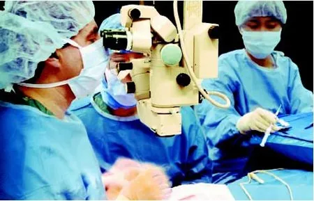
Reproductive surgeon and assistant working under the operating microscope
Microsurgical equipment greatly magnifies the operating field, provides instrumentation precise and delicate enough to maneuver under high magnification and allows the reproductive surgeon the ability to operate on structures barely visible to the human eye. A magnification of five to forty times (5-40x) is generally required for microsurgery. A lower magnification may be used to identify and expose structures, while a higher magnification is most often used for microsurgical repair. Gynecologic suturing usually requires gauges of 2-0 (0.3 mm) to 6-0 (0.07 mm). Conversely, gauges of 9-0 (0.03 mm) to 12-0 0.001 mm) are generally used for microsurgery. Dr. Lipskind uses 8-0 and 9-0 micro-sutures for microsurgical end to end an astomosis in reversing a tubal sterilization. The most important tools used by the microsurgeon are the microscope, microsurgical instruments, and micro-sutures.
Lighting is enhanced by maintaining a low level of light in the rest of the operating room, while turning up the light intensity of the operating microscope. The operating microscope used by Dr. Lipskind has two operative lens, so both he and his assistant have their own view with controlled magnification and focus. The microscope is wired to a high definition monitor, HD camera and HD printer, so the entire OR team can see what is going on. This makes the procedure go more efficiently, and allows the opportunity to record the entire procedure and/or take high quality HD pictures to be able to show the family exactly what was done.
For a reproductive surgeon to learn and perform microsurgery, extensive training and practice are required. Extensive and ongoing practice is necessary for a reproductive surgeon to maintain adequate proficiency at microsurgical techniques. If these procedures are only being done a few times per year, the surgeon will not be able to maintain his/her proficiency and expertise.
Dr. Lipskind received extensive experience and training in microsurgical surgeries and techniques and reversals of female sterilization and performs several microsurgeries per month. He has completed hundreds of tubal ligation reversals with very high rates of success. Those same microsurgical techniques are strictly followed in all his surgeries, whether they are by laparoscopy or during open cases (laparotomy). The goal is ALWAYS to preserve and enhance a woman's childbearing potential.


